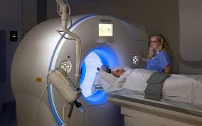
CT Scans and Risk to Health
I was motivated to write this researched article after many of my patients have been asking questions about how harmful are repetitive CT scans for diagnostic purposes. I have personally encountered patients who have had 8 – 10 CT scans in a period of only 3 – 4 years, and it is certain that there are many more in the same situation.
Hopefully this article will provide much needed information and scientific references to aid both patients and doctors alike in the sensible use of CT scans, as well as the use of radiation therapy as a cancer treatment.
One third of people getting a CT scan didn’t know the test exposed their body to radiation. Moreover, and more significant, 75% of an entire group of health professionals significantly underestimated the radiation dose from a CT scan, and 53% of radiologists and 91% of emergency-room physicians did not believe that CT scans increased the lifetime risk of cancer!
In a new study (http://archinte.jamanetwork.com/article.aspx?articleid=1487286) from a U.S. medical center, researchers found that 85% of patients also underestimated the amount of radiation delivered by a CT scan, and only 5% of them thought the scan would increase their chances of getting cancer again in their lifetime (1).
It is estimated that more than 62 million CT scans per year are currently obtained in the United States, including at least 4 million for children. (2)
CT scans are high-powered X-rays that expose patients to between ten and 100 times more radiation than a normal head or chest X-ray for example.
By its nature, CT involves larger radiation doses than the more common, conventional X-ray imaging procedures (Table 1).
The radiation dose from one CT scan typically ranges from a level comparable to yearly background radiation from natural sources (like the earth and sun) – to close to 20 millisieverts, which is the annual exposure limit for nuclear industry employees.
| Table 1. Typical Organ Radiation Doses from Various Radiologic Studies
|
||
| Study Type | Relevant Organ | Dose (mGy or mSv)
|
|
Dental radiography Posterior–anterior chest radiography Lateral chest radiography Screening mammography Adult abdominal CT with Barium enema Neonatal abdominal CT |
Brain Lung Lung Breast Stomach Colon
Stomach
|
0.005 0.01 0.15 3 10 15
20 |
Taken from: Computed Tomography — An Increasing Source of Radiation Exposure. David J. Brenner, Ph.D., D.Sc., and Eric J. Hall, D.Phil., D.Sc. N Engl J Med 357;22 www.nejm.org November 29, 2007.
MEASURING RADIATION EXPOSURE
The radiation dose is a measure of ionizing energy absorbed per unit of mass. Radiation exposure is measured in two units of measurement:
- The gray (Gy) – one gray is the absorption of one joule of energy from ionizing radiation, per kilogram of matter. A gray measures the actual absorbed dose to a specific area, like to your brain or prostate, for example. This can also be expressed as milligrays (mGy).
- The sievert (Sv) – One sievert is a measurement of the equivalent dose of total body exposure. Like comparing a dose to your prostate with the same dose equally spread across your whole body. The sievert is a more appropriate measurement for exposure of nuclear workers or bomb victims. This can also be expressed as millisieverts (mSv).
So what does all this mean in reality? Putting it into perspective, we can say that if your whole body was exposed to 5 gray of high-energy radiation in one blast you would be dead in about 14 days.
TYPICAL ORGAN RADIATION EXPOSURE
For example, a conventional anterior – posterior abdominal X-ray examination results in a dose to the stomach of approximately 0.25 mGy, which is at least 50 times smaller than the corresponding stomach dose from an abdominal CT scan (Table 2).
| Table 2. TYPICAL ORGAN CT-RADIATION EXPOSURE COMPARED TO X-RAYS
|
||
| BODY PART | RADIATION DOSE | COMPARABLE X-RAYS |
| Standard Chest X-ray | 0.01-0.15 mGy | 1 |
| Head CT | 56 mGy | 373 X-rays |
| Cardiac CT | 40-100 mGy | 266-666 X-rays |
| Mammogram | 3 mGy | 20 X-rays |
| Neonatal Abdominal CT | 20 mGy | 133 X-rays |
| Barium Enema | 15 mGy | 100 x-rays |
| Abdomen CT | 14 mGy | 93 x-rays |
| Chest CT | 13 mGy | 86 x-rays |
| Chest/Abd/Pelvis | 12 mGy | 80 x-rays |
RISKS ASSOCIATED WITH LOW-DOSE RADIATION
Ionizing radiation from CT scans is powerful enough to damage DNA. In biologic material exposed to X-rays, the most common scenario is the creation of hydroxyl radicals from x-ray interactions with water molecules; these radicals in turn interact with nearby DNA to cause strand breaks or base damage. X-rays can also ionize DNA directly. Most radiation-induced damage is rapidly repaired by various systems within the cell, but DNA double-strand breaks are less easily repaired, and occasional disrepair can lead to induction of gene fusions, all of which are linked to the induction of cancer. (3)
And if the DNA damage is not repairable by the body it can lead to cancer. Depending on the machine settings, the organ being studied typically receives a radiation dose in the range of 15 millisieverts (mSv) (in an adult) to 30 mSv (in a neonate) for a single CT scan, with an average of two to three CT scans per study. At these doses, as reviewed elsewhere, (4 – 5) the most likely (though small) risk is for radiation-induced carcinogenesis.
Most of the quantitative information that we have regarding the risks of radiation-induced cancer comes from studies of survivors of the atomic bombs dropped on Japan in 1945. (6) Data from cohorts of these survivors are generally used as the basis for predicting radiation-related risks in a population because the cohorts are large and have been intensively studied over a period of many decades, they were not selected for disease, all age groups are covered, and a substantial subcohort of about 25,000 survivors (7) received radiation doses similar to those of concern here – that is, less than 50 mSv.
Of course, the survivors of the atomic bombs were exposed to a fairly uniform dose of radiation throughout the body, whereas CT involves highly non-uniform exposure, but there is little evidence that the risks for a specific organ are substantially influenced by exposure of other organs to radiation. There was a significant increase in the overall risk of cancer in the subgroup of atomic-bomb survivors who received low doses of radiation, ranging from 5 to 150 mSv (6 – 8); the mean dose in this subgroup was about 40 mSv, which approximates the relevant organ dose from a typical CT study involving two or three scans in an adult.
Although most of the quantitative estimates of the radiation-induced cancer risk are derived from analyses of atomic-bomb survivors, there are other supporting studies, including a recent large-scale study of 400,000 radiation workers in the nuclear industry (9 – 11) who were exposed to an average dose of approximately 20 mSv (a typical organ dose from a single CT scan for an adult).
A significant association was reported between the radiation dose and mortality from cancer in this cohort (with a significant increase in the risk of cancer among workers who received doses between 5 and 150 mSv); the risks were quantitatively consistent with those reported for atomic bomb survivors.
CT SCANS AND CANCER
Since the inception of CT in the 1970s, its use has increased rapidly. It is estimated that more than 62 million CT scans per year are currently obtained in the United States, including at least 4 million for children. (12).
One study from the National Cancer Institute estimated there would be about 29,000 future cancers related to scans done in 2007 alone.
The most common cancers thought to be caused by radiation exposure are lung, breast, thyroid, stomach and leukemia.
RADIATION THERAPY FOR CANCER PATIENTS
The radiation exposure for cancer patients undergoing radiation therapy is obviously a lot more than for diagnostic CT scans.
Exposure from radiation therapy ranges from 20 – 80 gray depending on the type of cancer. In the example above we mentioned that a whole body exposure of 5 gray would kill you in two weeks. In the case of radiation therapy, as it is very concentrated on one area of the body, usually the tumour, then it does not kill you in a short period of time as the whole body is not exposed.
SIDE EFFECTS OF RADIATION THERAPY
Many will know that radiation therapy can result in hair loss – indeed, 1 gray will cause hair loss in the radiated area, whereas over 10 gray would burn the hair follicles making the hair loss permanent.
Saliva glands, tear glands, sweat glands and vaginal mucosa glands can be permanently damaged by radiation over 30 gray, leading to chronic dry mouth, dry eyes, dry skin
and dry “other parts” for the rest of your life.
Around 8 gray to the ovaries causes permanent infertility.
IGNORANCE AMONGST HEALTH PROFESSIONALS
Why are CT scans still used by doctors at such a high frequency, when the risks of developing cancer and other serious side-effects are real?
Part of the issue is that physicians often view CT studies in the same light as other radiologic procedures, even though radiation doses are typically much higher with CT than with other radiologic procedures.
In a recent survey of radiologists and emergency-room physicians, (13) about 75% of the entire group significantly underestimated the radiation dose from a CT scan, and 53% of radiologists and 91% of emergency-room physicians did not believe that CT scans increased the lifetime risk of cancer.
In the light of these findings, the pamphlet “Radiation Risks and Pediatric Computed Tomography (CT): A Guide for Health Care Providers,” (14) which was recently circulated among the medical community by the National Cancer Institute and the Society for Pediatric Radiology, is most welcome.
In summary, there is direct evidence from epidemiologic studies that the organ doses corresponding to a common CT study (two or three scans, resulting in a dose in the range of 30 to 90 mSv) result in an increased risk of cancer. The evidence is reasonably convincing for adults and very convincing for children.
When it is estimated that only in America there are about 70 million CT scans run annually, at about $500 per scan, then one can quickly see that this is a multi-billion dollar industry (70 million CT scans at $500 each equals 35 billion dollars)!
Dr. Amy Berrington de Gonzalez has spent the last 15 years studying medical radiation exposure. She made a presentation of her findings at the NIH on January 11, 2013 entitled, “Medical Radiation and Cancer Risk: Assessing the Price of Progress” – http://videocast.nih.gov/summary.asp?Live=12262&bhcp=1
Dr de Gonzalez concluded the following in her presentation:
Medical radiation is one of the top 10 carcinogens in the USA – here are the figures for the top 10 causes of cancer in the US:
1) Smoking 20%
2) Diet 10%
3) Obesity 6%
4) Alcohol 5%
5) Infections 3%
6) Medical Radiation 1-3%
7) Physical Inactivity 1%
8) Post-menopausal Hormone Drugs .05%
Currently diagnostic radiation (CT Scans, etc.) is attributed to causing 1% of all cancers. Dr. Berrington projected that if we keep using diagnostic radiation at the current levels, the number of radiation related cancers will triple in the coming years.
She also states that radiation treatments are in the top five causes of secondary cancers for cancer survivors.
On average, cancer survivors have a 14% risk of a recurrence, which researchers attribute to three factors: lifestyle, treatments, and genetics. (We know from the field of epigenetics that gene expression is influenced by your lifestyle and toxic exposure including cancer treatments. In other words, your genetics do not ultimately determine your fate – your diet and lifestyle choices play a significant role in your fate.)
8% of secondary cancers (on average) are related to radiation therapy. The percentage is a bit higher for prostate (11%), cervix (18%) and testicular (25%).
This 8% figure only represents what scientists can statistically prove, with the likelihood that it will be much higher.
After only one CT Scan, the risk of radiation related cancer remains elevated throughout the rest of your life. Dr. Berrington estimates that the risk is 1 per 10,000 to develop cancer within 10 years and 1 per 1,000 to develop cancer in their lifetime.
So one CT Scan doesn’t seem very risky, but keep in mind that the risk increases significantly with every scan.
Also it should be noted that the risk of cancer from CT Scans is much higher for babies, children and young adults. The risk decreases significantly after age 25 and stays consistent from there to age 80.
Dr. Berrington also mentioned that the new backscatter X-rays used in airports are 10,000 times weaker than a standard X-ray.
RADIATION MAKES CANCER MORE AGGRESSIVE
Researchers from the Department of Radiation Oncology at the UCLA Jonsson Comprehensive Cancer Center report that radiation treatment transforms cancer cells into treatment-resistant breast cancer stem cells, even as it kills half of all tumor cells (15).
In this study published in the July 2012 Cancer Journal (16), researchers from the Department of Radiation Oncology at UCLA reported that radiation treatment actually drives breast cancer cells into greater malignancy.
They found that even when radiation kills half of the tumor cells treated, the surviving breast cancer stem cells (iBCSCs), were up to 30 times more likely to form tumors than the non-irradiated breast cancer cells. And they became resistant to further treatments.
Their findings show that if tumors are challenged by certain stressors that threaten them (such as radiation), they generate iBCSCs that may, along with surviving cancer stem cells, produce more tumors.
When radiation shrinks a tumor everyone thinks it’s working, but that can be very misleading. What may be happening is that the radiation is only increasing the ratio of highly malignant cells to benign cells inside the tumor, which reproduce rapidly.
Many of the cells in a tumor are not actually cancerous. Destroying those benign cells with radiation will shrink the size of the tumor but not necessarily affect the cancerous cells.
There was another similar study published in 2012 in the journal Stem Cells (17) entitled, “Radiation-induced reprogramming of breast cells”.
In this study researchers found that ionizing radiation reprogrammed less malignant (more differentiated) breast cancer cells into iBCSCs (breast cancer stem cells).
Basically radiation creates cancer super cells, which are treatment resistant. Kind of like the superbugs you hear about in hospitals that are resistant to antibiotics.
And this study entitled: “Long-term recovery of irradiated prostate cancer increases cancer stem cells” (18) showed that radiotherapy increases cancer stem cells in the prostate, ultimately resulting in cancer recurrence and worsened prognosis.
WHAT CAN WE DO?
It seems that about one third of all CT scans are not justified by medical need, and it appears to be likely, (1) perhaps 20 million adults and, crucially, more than 1 million children per year in the United States are being irradiated unnecessarily.
Perhaps other less harmful forms of imaging should be utilized diagnostically, such as ultrasound scans as well as Magnetic Resonance Imaging (MRI) scans.
In addition, many of the parameters that actually increase the radiation dosage are under the control of the radiologist who should always lean on the side of caution and give the absolute minimum dose of radiation to the patient.
REFERENCES
1. Janet M. Busey, MS; Laurie A. Soine, PhD, ARNP; Jenine R. Yager, BA; Eunice Choi, BS; William P. Shuman, MD. Patient Knowledge and Understanding of Radiation From Diagnostic Imaging. Arch Intern Med. 2012;():1-2. doi:10.1001/2013.jamainternmed.1013.
2. What’s NEXT? Nationwide Evaluation of X-ray Trends: 2000 computed tomography. (CRCPD publication no. NEXT_2000CTT.) Conference of Radiation Control Program Directors, Department of Health and Human Services, 2006. (Accessed November 5, 2007, at http://www.crcpd.org/Pubs/NexTrifolds/NEXT2000CT.pdf
3. Mitelman F, Johansson B, Mertens FE. Mitelman. Database of chromosome aberrations in cancer. Cancer Genome Anatomy Project, 2007. (Accessed November 5, 2007
4. Brenner DJ, Doll R, Goodhead DT, et al. Cancer risks attributable to low doses of ionizing radiation: assessing what we really know. Proc Natl Acad Sci U S A 2003;100:13761-6.
5. Health risks from exposure to low levels of ionizing radiation – BEIR VII. Washington, DC: National Academies Press, 2005.
6. Preston DL, Pierce DA, Shimizu Y, et al. Effect of recent changes in atomic bomb survivor dosimetry on cancer mortality risk estimates. Radiat Res 2004;162:377 – 89.
7. Preston DL, Shimizu Y, Pierce DA, Suyama A, Mabuchi K. Studies of mortality of atomic bomb survivors. Report 13: Solid cancer and non-cancer disease mortality: 1950-1997. Radiat Res 2003;160:381- 407.
8. Pierce DA, Preston DL. Radiation related cancer risks at low doses among atomic bomb survivors. Radiat Res 2000; 154:178-86.
9. Preston DL, Ron E, Tokuoka S, et al. Solid cancer incidence in atomic bomb survivors: 1958-1998. Radiat Res 2007;168: 1-64.
10. Cardis E, Vrijheid M, Blettner M, et al. The 15-country collaborative study of cancer risk among radiation workers in the nuclear industry: estimates of radiation-related cancer risks. Radiat Res 2007;167: 396-416.
11. Idem. Risk of cancer after low doses of ionising radiation: retrospective cohort study in 15 countries. BMJ 2005;331:77.
12. What’s NEXT? Nationwide Evaluation of X-ray Trends: 2000 computed tomography. (CRCPD publication no. NEXT_2000CTT.) Conference of Radiation Control Program Directors, Department of Health and Human Services, 2006. (Accessed November 5, 2007.
13. Lee CI, Haims AH, Monico EP, Brink JA, Forman HP. Diagnostic CT scans: Assessment of patient, physician, and radiologist awareness of radiation dose and possible risks. Radiology 2004;231:393-8.
14. Radiation risks and pediatric computed tomography (CT): a guide for health care providers. Rockville, MD: National Cancer Institute. (Accessed November 5, 2007, at http://www.nci.nih.gov/cancertopics/causes/radiation-risks-pediatric-CT.)
15. Slovis TL, Berdon WE. Panel discussion. Pediatr Radiol 2002;32:242-4.
16. Lagadec, C, Vlashi, E, Della Donna, L, Dekmezian, C, Pajonk, F. Radiation-induced reprogramming of breast cancer cells. Stem Cells. 2012;30:833–844.
17. Carrie Printz. Radiation treatment generates therapy-resistant cancer stem cells from less aggressive breast cancer cells. Cancer. Volume 118, Issue 13,page 3225, 1 July 2012
18. Cho YM, Kim YS, Kang MJ, Farrar WL, Hurt EM. Long-term recovery of irradiated prostate cancer increases cancer stem cells. Prostate. 2012 Dec 1;72(16):1746-56. doi: 10.1002/pros.22527. Epub 2012 Apr 18.
Researched and written by:
Dr George J Georgiou, Ph.D.,N.D.,DSc (AM).,MSc.,BSc
Holistic Health Practitioner
Director, Da Vinci Holistic Health Center
Larnaca, Cyprus
www.naturaltherapycenter.com